OnGoing Project:
snake fungal disease
|
What is Snake Fungal Disease (SFD)?
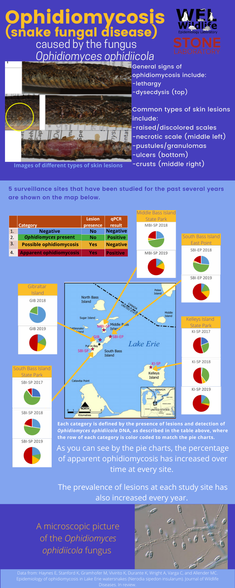
Snake Fungal Disease (SFD) is an emerging disease caused by the fungal pathogen Ophidiomyces ophiodiicola. When a snake has SFD, it often displays symptoms of blisters, crusty sores, thickened scales, pustules and facial disfiguration. When did SFD emerge? SFD emerged in 2000 and LEWS were one of the original eleven species of snakes who were impacted by the disease. Does SFD look the same in all snake species? No! SFD affects every species of snake differently! There are more than 40 species of snake that have been accepted. In vipers (i.e. cottonmouths, rattlesnakes, etc.), Ophidiomyces stays on and impacts only the surface of the skin. In colubrids (i.e. watersnakes, garter snakes, etc.), Ophidiomyces can go deeper than the surface of the skin, impacting muscles, bones, and organs, too. Even within a species, symptoms can vary widely. How is Ophidiomyces transmitted? Ophidiomyces is a fungus that can tolerate a wide range of soil types, pH, and temperatures. With that being said, this fungus is very resilient in the environment and can survive in a wide variety of habitats. However, there is currently no evidence showing that Ophidiomyces is transmitted through the environment. Instead, current research shows that it is spread via contact with animals who have the fungus on their skin. In September 2019, a new study was conducted at Stone Laboratory to find out whether or not LEWS mothers who tested positive for SFD were transmitting the fungus to their offspring during birth. Although we are still waiting on the results of this study, we are excited to find out the answer to this aspect of SFD epidemiology! How do you diagnose SFD? There is no perfect picture of SFD. Unfortunately, there is no quick and easy way to diagnose SFD. This disease displays itself in a wide variety of ways that could also be indicative of other diseases that impact reptiles. Diagnosis can only be made by extensively swabbing affected snakes. Samples on a cotton swab are processed via culturing or qPCR in a lab, and only afterwards can a diagnosis be made. It is possible to get false negative results from this test, but false positive results do not exist. To be considered a positive case of Ophidiomycosis the snake will have a positive qPCR result and display visible symptoms of the fungus. What is the prognosis for LEWS with SFD? Short answer: We don't know yet. Team Snake started swabbing LEWS for SFD in May 2017. Of 252 swabs from 181 individuals taken throughout the summer of 2017, 75.4% tested positive for the presence of the fungus. However, presence of the fungus does not mean the snake is infected with SFD. It is thought that some snakes are able to avoid infection from Ophidiomyces but still harbor the fungus on their skin, which can then be transmitted to other snakes in the population. We are continuing to monitor our study sites every year to determine the prevalence of this disease among the LEWS populations, with over 300 swabs collected in 2019. We are are also hoping that this monitoring will allow us to track the health of individuals over time to better understand the progression of the disease in the wild. Check back here soon for updates from current research! What are we doing to further this research? By working closely with the wildlife epidemiology lab at the University of Illinois and continuing to take swabs from Lake Erie watersnakes, we hope to learn more about this fungus and answer questions such as: Once a snake tests positive, will it always be infected? Are there environmental factors that are affecting the fungal load? Will the fungal stay at the same level over time or gradually get worse? What can you do to help? If you see a snake in the wild that you think may be suffering from this disease, contact and alert your local state wildlife agency. Always follow all laws and regulations regarding interacting with wildlife! If you are interacting with wildlife on a regular basis, please do your part to prevent the spread of this disease! Wear gloves and disinfect your equipment using solutions that inhibit the growth of Ophidiomyces, such as bleach or another approved solution. Additionally, cleaning your shoes/attire when traveling to different wildlife areas can help stop the spread of this fungus. To learn more about other effective disinfectants, click here! The information on this page was obtained from Dr. Matthew C. Allender at the Wildlife Epidemiology Laboratory at the University of Illinois. |
Facial disfiguration caused by SFD
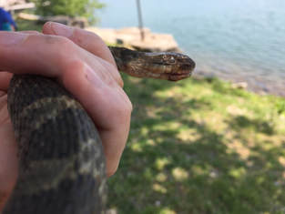
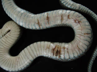
Blisters caused by SFD
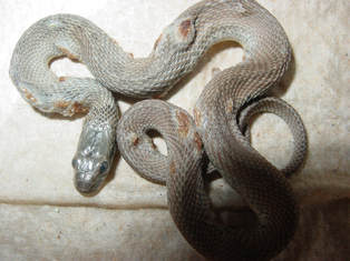
Current known US distribution of
O. ophidiicola (Allender et al. 2015) 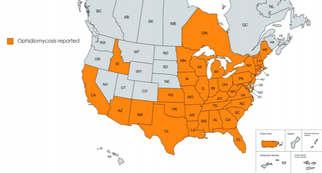
US reports of O. ophidiicola (Haynes 2020)
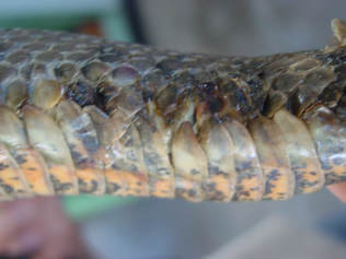
Crusty sores/ulcers caused by SFD
Crusty sores/ulcers caused by SFD
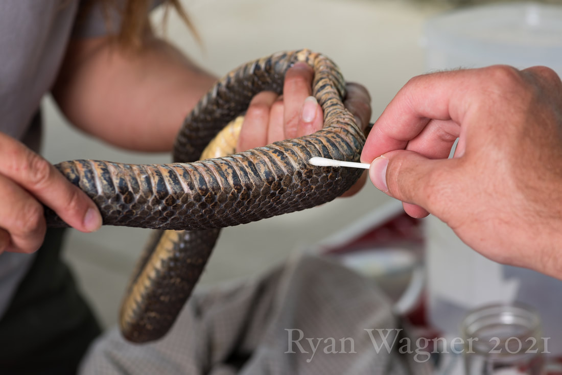
Swabbing of a snake focusing on ventral lesions
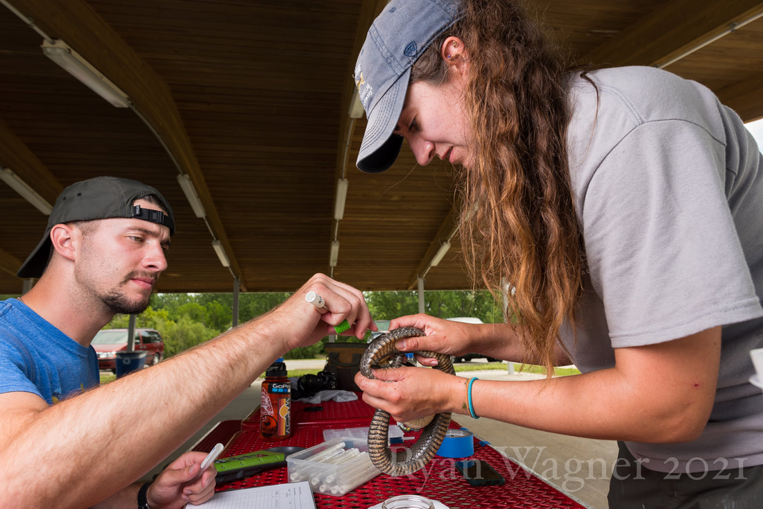
After completing a full body swab we will return to focus on symptomatic areas on the body
|